Sleep is divided into several stages, each with distinct patterns of brain activity.
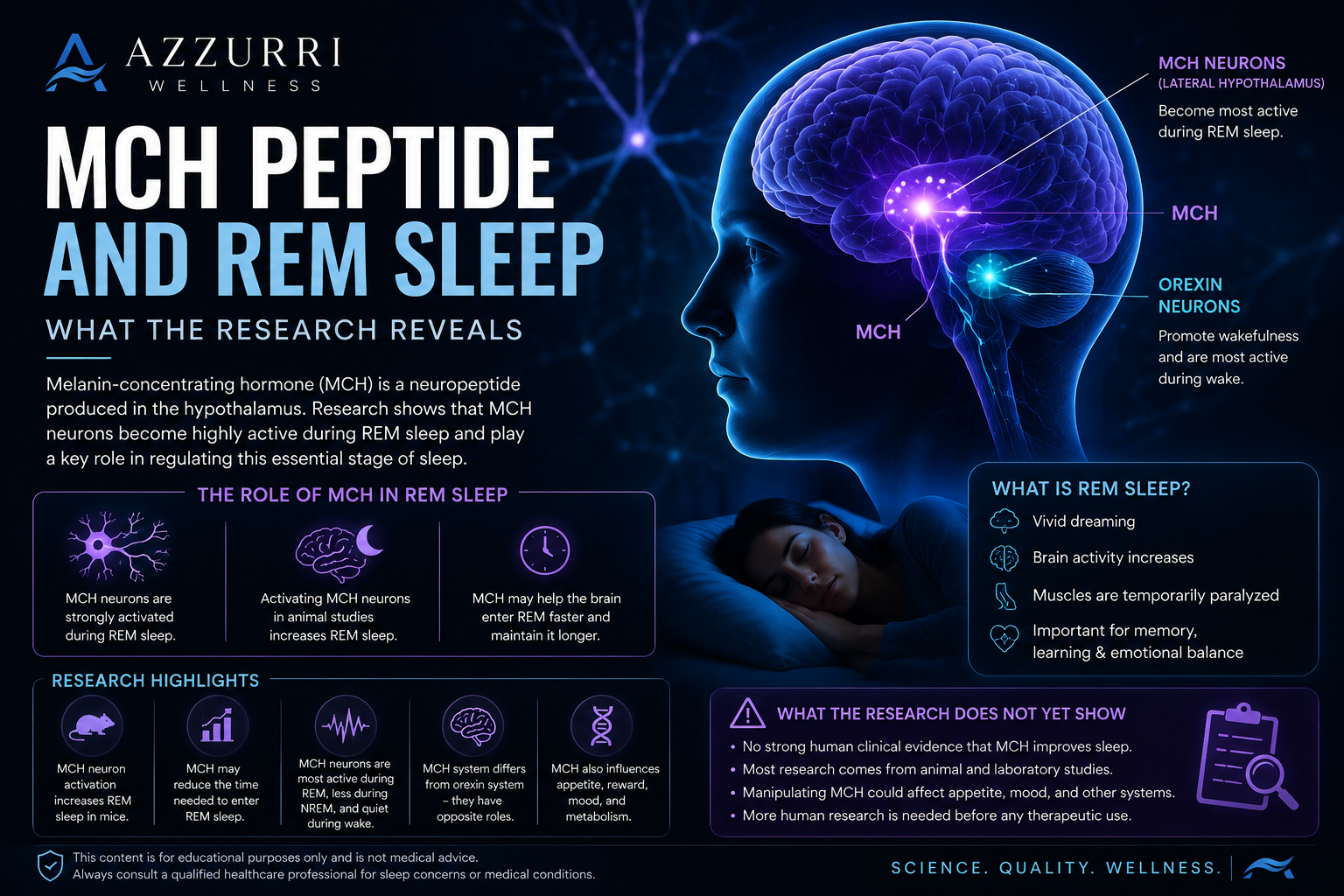
Deep non-REM sleep is commonly associated with slow brain waves and physical restoration. Rapid eye movement sleep, or REM sleep, is characterized by vivid dreaming, increased brain activity, temporary muscle paralysis, and processes connected with memory and emotional regulation.
One peptide system receiving attention in REM research is melanin-concentrating hormone, or MCH.
Despite its name, MCH is not the same as melanin, the pigment responsible for skin and hair color. MCH is a neuropeptide produced by a specific group of neurons in the hypothalamus. Research indicates that these neurons become particularly active during REM sleep.
What Is Melanin-Concentrating Hormone?
MCH is a peptide produced mainly by neurons in the lateral hypothalamus.
This brain area contains several populations of neurons involved in sleep, wakefulness, appetite, reward, and energy regulation. MCH neurons project widely throughout the brain, allowing them to influence multiple systems.
Early research focused heavily on MCH and feeding behavior. More recent studies have established that the same neural population also participates in sleep-wake regulation, especially REM sleep.
MCH and Orexin Have Different Roles
MCH neurons are located near neurons that produce orexin, also known as hypocretin.
Although they occupy neighboring areas, the systems generally show contrasting patterns:
- Orexin neurons are strongly associated with wakefulness and stable alertness.
- MCH neurons are largely inactive during active waking and become more active during sleep, particularly REM sleep.
This does not mean they operate as simple on-and-off switches. Sleep regulation involves many overlapping neurotransmitters and neural circuits. Still, their contrasting activity illustrates how the hypothalamus helps shift the brain between wakefulness and different sleep stages.
Why REM Sleep Matters
REM sleep occurs several times throughout the night, with longer REM periods generally appearing closer to morning.
During REM sleep:
- Brain activity becomes more similar to waking activity.
- Most skeletal muscles become temporarily inhibited.
- Rapid eye movements occur.
- Dreaming is often vivid.
- Emotional and memory-processing networks remain active.
REM sleep is not automatically better than non-REM sleep. Healthy sleep requires an organized sequence of both.
Increasing one stage without understanding the effects on the others would not necessarily improve overall sleep quality.
What Happens to MCH Neurons During Sleep?
Recording studies indicate that MCH neurons are most active during REM sleep, less consistently active during non-REM sleep, and generally quiet during waking.
This activity pattern led researchers to investigate whether MCH neurons merely become active during REM sleep or actively help produce and maintain it.
Reviews of the field conclude that MCH signaling forms an important component of sleep-wake circuitry, with particularly strong evidence connecting it to REM regulation.
What Activation Studies Found
Modern neuroscience allows researchers to selectively activate or inhibit specific populations of neurons.
In one mouse study, chemogenetic activation of MCH neurons produced a substantial increase in REM sleep without significantly increasing wakefulness or non-REM sleep. Researchers concluded that activation of the MCH system primarily drove REM sleep under those experimental conditions.
Another study found that MCH neurons could promote REM sleep during normal physiological conditions and may support quicker transitions into REM.
Earlier experiments involving MCH administration also reported increases in REM sleep and reductions in the time required to enter REM sleep. However, direct administration may expose the brain to concentrations that differ from natural signaling.
Results from those experiments must therefore be interpreted cautiously.
Why Different Studies Sometimes Disagree
Not every MCH experiment has produced identical findings.
Some studies observed increases in both non-REM and REM sleep, while others found changes primarily in REM sleep. These differences may reflect:
- The method used to activate neurons
- The duration and frequency of stimulation
- The time of day
- The animal model
- Whether the peptide, receptor, or entire neuron population was manipulated
- Whether activation occurred during wakefulness or an existing sleep stage
Artificially stimulating a neuron for several hours is not the same as reproducing its natural firing pattern.
This is one reason researchers avoid reducing sleep regulation to a single peptide.
MCH, Dreaming, and Memory
Because MCH neurons are active during REM sleep, researchers have also investigated possible connections with memory processing.
Some MCH neurons project into the hippocampus and other areas involved in learning and memory. Experimental work suggests that MCH-related activity during sleep may influence which information is retained or weakened.
However, this remains an evolving area of neuroscience. It would be premature to claim that MCH improves memory, controls dreaming, or produces emotional recovery in humans.
The relationship between REM sleep and memory is itself complex. Different types of learning may rely on different sleep stages, and memory processing involves many brain regions and signaling systems.
Does MCH Offer a Proven Way to Improve Sleep?
No established human clinical evidence currently shows that MCH administration safely improves routine sleep quality or treats insomnia.
Most direct evidence comes from:
- Animal experiments
- Neural recording studies
- Chemogenetic or optogenetic manipulation
- Administration directly into the central nervous system
- Receptor studies performed under controlled laboratory conditions
These approaches are valuable for identifying mechanisms. They do not provide instructions for consumer use.
MCH also participates in appetite, metabolism, reward, and mood-related pathways. Manipulating the system could therefore produce effects extending well beyond sleep.
REM Sleep Is Only One Part of Healthy Sleep
A normal night includes repeated cycles of:
- Light non-REM sleep
- Deeper slow-wave sleep
- REM sleep
Sleep quality cannot be judged by maximizing a single stage.
For example, frequent awakenings may fragment REM sleep even when total time in bed is long. Sleep apnea can repeatedly interrupt normal sleep architecture. Certain medications and substances can also alter REM timing.
A complete sleep assessment must therefore consider:
- Total sleep duration
- Sleep onset
- Number of awakenings
- Breathing patterns
- Daytime alertness
- Circadian timing
- Balance among sleep stages
Why the MCH Research Still Matters
MCH research reveals that sleep stages are regulated by dedicated networks rather than emerging passively when the brain becomes inactive.
It also demonstrates that the hypothalamus contains neighboring systems capable of supporting very different states. Orexin-related pathways stabilize wakefulness, while MCH-related activity appears to facilitate REM sleep.
Understanding this balance may eventually contribute to research on insomnia, hypersomnia, mood disorders, memory, and REM-related conditions.
However, scientific interest should not be confused with established clinical application.
Final Takeaway
Melanin-concentrating hormone is one of the most interesting peptide systems involved in REM sleep regulation.
Animal and neural-circuit studies indicate that MCH neurons become highly active during REM sleep and that experimental activation can increase REM duration or facilitate entry into REM.
The findings improve scientific understanding of sleep architecture, but direct human evidence remains insufficient to describe MCH as a proven sleep-improvement treatment.
Research References
- Bandaru SS, et al. Sleep-wake control by melanin-concentrating hormone neurons. View research
- Torterolo P, et al. Melanin-concentrating hormone and its role in REM sleep. View research
- Vetrivelan R, et al. Melanin-concentrating hormone neurons specifically promote REM sleep in mice. View research
- Naganuma F, et al. MCH neurons contribute to the induction and maintenance of REM sleep. View research
- Torterolo P, et al. Melanin-concentrating hormone: a new sleep factor? View research
- Potter LE, et al. The melanin-concentrating hormone system as a research target. View research